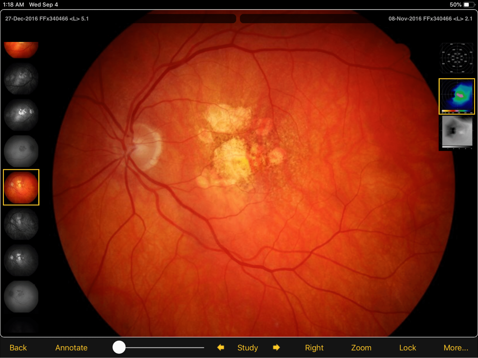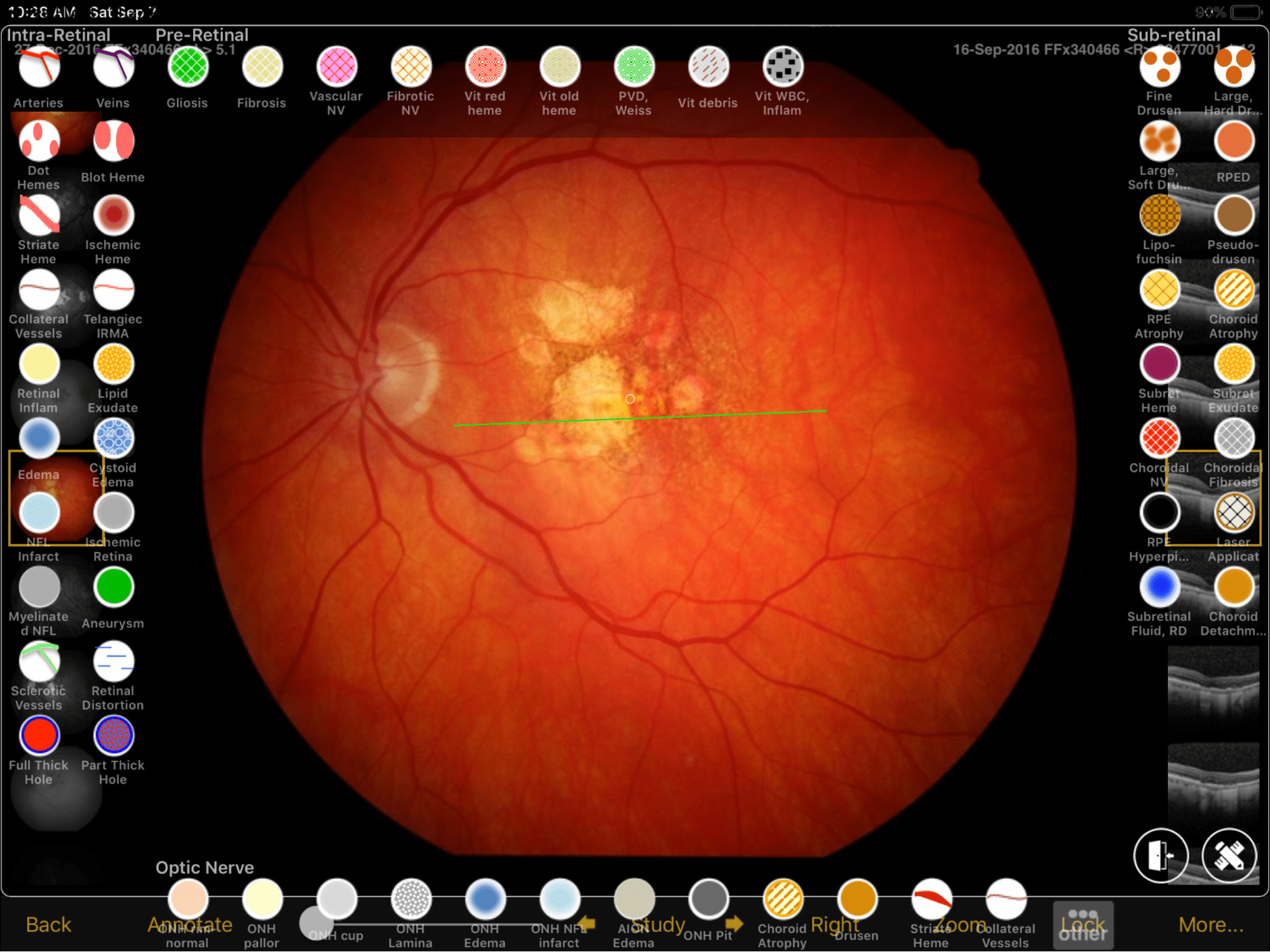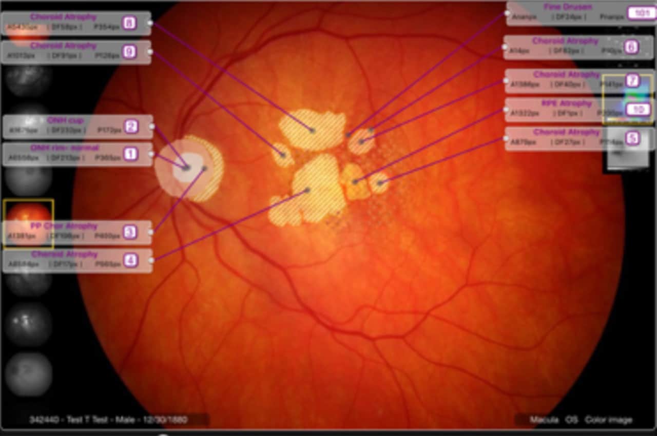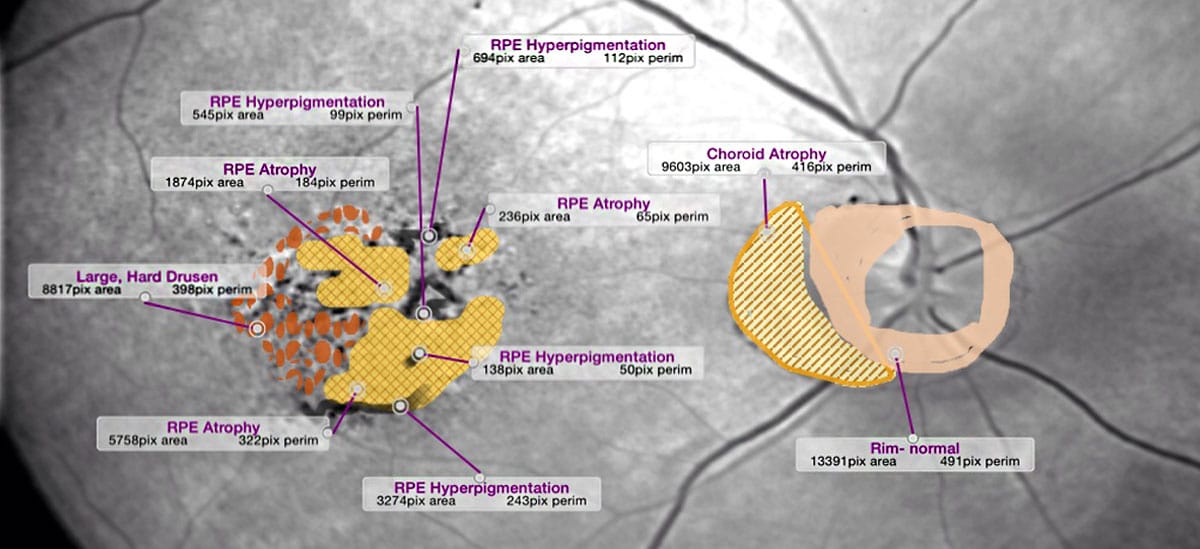
Physician drawings have represented a method for documenting features noted on retinal examination for as long as clinicians have examined the fund us. Although the understanding of both posterior and peripheral retinal pathology has evolved considerably, the method of documentation by retinal drawing has essentially not changed from the early concepts. Although clinicians are moving away from paper charts to electronic medical records, the method to provide drawing tools has remained a palette of colors. This requires the clinician not only to draw the lesions he desires, but then he must label the lesions drawn and then in the examination findings he must repeat what he has observed and drawn. Furthermore, with the availability of retinal images, physicians are limited in their documentation to descriptions of pathology with poor correspondence between examinations and between viewers.
Sinclair Technologies has developed Ocudraw, a formalized system for coding pathology by allowing the viewer to draw his findings using a template of pathologic lesions such that the position and dimensions are digitally quantitated that provides improved follow up comparison and management and to insure more comprehensive communication among providers. With Ocudraw Lite, the physician draws his observations on examination using a palette of pathologic lesions. Subretinal lesions are drawn beneath intra-retinal with pre-retinal lesions placed on top either on templates of the posterior retina or those of the full extent of the retina. When the drawing is saved, the pathology is identified in the image with the lesions contained in the DICOM header for inclusion as findings in electronic charts. Ocudraw Pro provides all of this including as well the ability to draw over imported retinal or OCT images, with the additional capability of defining areas and perimeters of the pathology identified. In this caseDICOM compliant image header can be extracted for inclusion in the electronic medical record and research databases where they can be reviewed in a “crowd sourced” image management system, that will effectively allow for the development of quantitated methods for image management of disease progression, something badly needed to improve predictions of outcomes.


